The Microscope | Teacher Chello
Chello Gregorio
Overview
This video provides a comprehensive introduction to the microscope, covering its history, types, parts, and usage. It begins by explaining the fundamental purpose of microscopy in revealing the unseen world and traces the evolution of the microscope from simple lenses to sophisticated compound and electron microscopes. The lecture details the optical and mechanical components of a light microscope, emphasizing the principles of magnification and resolution. It also briefly touches upon other scientific research tools like centrifuges and spectrophotometers, and concludes with essential guidelines for microscope care and handling. The overarching theme is the transformative impact of microscopy on scientific discovery.
Save this permanently with flashcards, quizzes, and AI chat
Chapters
- Microscopy is the scientific technique of using microscopes to view objects that are too small to be seen with the naked eye.
- The invention of the microscope allowed scientists to observe microstructures, leading to discoveries about organisms and molecules.
- Microscopes are essential tools in scientific research, enabling detailed observation and experimentation.
- The session aims to cover microscope parts, functions, types, usage, care, and other scientific research tools.
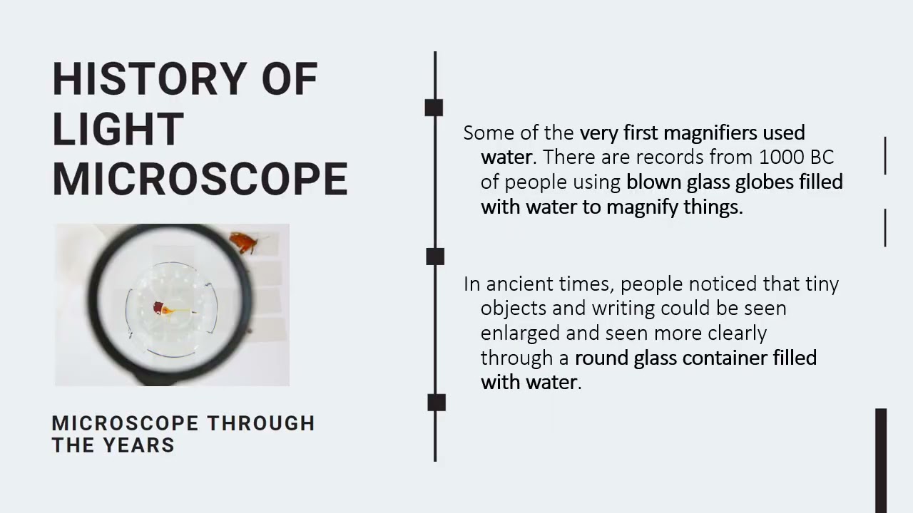
- Early magnifiers used water drops or simple lenses, with the first compound microscope appearing around 1590.
- The invention of spectacles around 1280-1285 in Italy paved the way for combining lenses.
- Zacharias Janssen is often credited with creating the first compound microscope around 1590 by combining two lenses.
- Robert Hooke improved the three-lens system, and his book 'Micrographia' documented microscopic observations, including the naming of 'cells'.
- Antonie van Leeuwenhoek developed highly effective single-lens microscopes, making groundbreaking observations of bacteria and protozoa.
- Magnification is the measure of how much larger a microscope makes an object appear, calculated by multiplying the objective lens magnification by the ocular lens magnification.
- Resolution is the smallest distance by which two points can be separated and still be distinguished as separate objects; higher resolution means better clarity and detail.
- The unit of measurement commonly used in microscopy is the micron (µm).
- Early microscopes produced large but blurry images, while modern advancements have led to large and clear images due to improved resolution.
- Light microscopes use visible light that passes through the specimen and is bent by lenses to produce a magnified image.
- They are suitable for observing living cells and processes, such as those found in a hay infusion.
- Different types of light microscopy exist, including bright-field, phase-contrast, dark-field, and fluorescence microscopy, each offering unique ways to visualize specimens.
- Confocal scanning laser microscopes use a concentrated beam of light to create detailed 3D views.
- Mechanical parts include the base, pillar, arm, stage, body tube, and adjustment knobs (coarse and fine).
- Optical parts include the eyepiece (ocular lens) and objective lenses (low power, high power, oil immersion).
- Illuminating parts provide light, such as a mirror or electric lamp, and are controlled by the substage condenser and iris diaphragm.
- The total magnification is the product of the ocular lens magnification and the objective lens magnification.
- Electron microscopes use a beam of electrons instead of visible light, offering much higher resolution and magnification.
- They have shorter wavelengths than visible light, allowing for the visualization of much smaller structures.
- Transmission Electron Microscopes (TEM) provide detailed internal views of cells, while Scanning Electron Microscopes (SEM) show surface details.
- Electron microscopes are significantly more expensive and complex than light microscopes.
- Proper handling involves carrying the microscope with two hands (one on the arm, one on the base) and placing it gently on the table.
- Preparation for use typically starts with the low power objective, followed by adjusting light and focusing.
- Maintenance includes cleaning lenses, storing the microscope properly, and seeking repairs when needed.
- Other scientific tools like centrifuges (for separating particles), chromatography (for separating pigments), electrophoresis (for separating substances via electric current), and spectrophotometers (for measuring light absorption) are also used in research.
Key takeaways
- The microscope is a fundamental tool that revolutionized our understanding of the biological world by revealing structures invisible to the naked eye.
- The development of the microscope has been a gradual process, with key innovations in lens technology and design.
- Magnification allows us to see objects larger, while resolution determines the clarity and detail of the magnified image.
- Light microscopes are versatile for observing living organisms and cellular processes, with various types offering different contrast methods.
- Electron microscopes provide significantly higher resolution, enabling the study of subcellular structures and viruses.
- Proper handling, care, and maintenance are crucial for the effective and long-term use of any microscope.
- A variety of other scientific instruments complement microscopy by enabling separation, analysis, and quantification of biological and chemical substances.
Key terms
Test your understanding
- What is the primary difference between magnification and resolution in microscopy?
- How did the invention of the spectacles influence the development of the compound microscope?
- Why is it important to start focusing with the low power objective lens when using a light microscope?
- What are the main advantages of using an electron microscope compared to a light microscope?
- Describe the proper procedure for carrying and setting up a microscope on a laboratory table.