13:24
Flow Cytometry Analysis
SENS Research Foundation
Overview
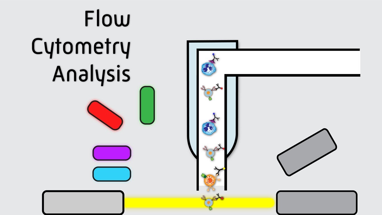
This video explains the principles and applications of flow cytometry, a powerful technique used to analyze individual cells within a population. It uses an analogy of a grocery store scanner to illustrate how cells are 'barcoded' with fluorescent antibodies and then passed through a laser beam. The laser detects signals from these tags, providing data on cell size, granularity, and the presence of specific molecules on the cell surface or inside. This allows researchers and clinicians to identify, quantify, and understand the function of different cell types, aiding in disease diagnosis and biological research.
How was this?
Save this permanently with flashcards, quizzes, and AI chat
Chapters
- Flow cytometry is a technique to analyze individual cells and cell populations.
- It helps understand cell function, health, and activity.
- It's analogous to a grocery store scanner that identifies items by barcode and weight.
Understanding flow cytometry is crucial for anyone in biological or medical fields, as it's a fundamental tool for cell analysis and discovery.
A grocery store scanner uses lasers to read barcodes and determine item identity and weight, similar to how a flow cytometer uses lasers to analyze cells.
- Cells are 'barcoded' using antibodies that specifically bind to unique molecules on or within the cell.
- Antibodies act like unique tags, allowing the flow cytometer to distinguish between different cell types.
- Careful selection of specific antibodies is essential to avoid misidentification of cells.
The specificity of antibody tagging directly determines the accuracy and resolution of the cell analysis, enabling precise identification of cell populations.
Using antibodies specific to proteins found on T-cells, NK cells, and neutrophils to differentiate these immune cell types within a blood sample.
- Fluorophores are molecules that absorb light and emit a detectable color.
- Antibodies are often linked to fluorophores, allowing them to be detected by the flow cytometer's lasers.
- Using different colored fluorophores is necessary when multiple antibodies are used in a single sample to distinguish between them.
Fluorophores are the 'detectors' that make the cellular barcodes visible, enabling the flow cytometer to gather information about the tagged molecules.
Linking a red-fluorescent molecule to an antibody that targets T-cells, so that T-cells will emit a red signal when excited by the laser.
- Cells are processed one at a time through a stream of fluid to ensure individual analysis.
- As each cell passes through a laser beam, it generates signals.
- Forward scatter (FSC) indicates cell size, and side scatter (SSC) indicates internal granularity or complexity.
The precise, single-cell interrogation by the laser is what allows flow cytometry to provide detailed information about each cell's physical characteristics.
A stream of cells flowing one by one into a laser beam, similar to items moving on a conveyor belt past a scanner.
- Detectors measure the scattered light (FSC, SSC) and fluorescence emitted by fluorophores.
- Data is typically visualized on plots, with FSC on the x-axis and SSC on the y-axis, showing clusters of different cell types.
- Fluorescence intensity plots show the number of cells versus the brightness of a specific fluorescent signal, indicating the expression level of the targeted molecule.
Interpreting these scatter and fluorescence plots is key to identifying and quantifying different cell populations based on their physical properties and molecular markers.
A scatter plot where dots cluster together, representing groups of cells with similar sizes (FSC) and granularity (SSC), allowing visual differentiation of cell types.
- Flow cytometry can analyze multiple markers simultaneously for more specific cell population separation.
- It's used in clinical settings for diagnosing diseases and in research for testing hypotheses.
- The technique provides insights into cell behavior and responses to environmental changes.
The ability to analyze cells at a detailed level has broad implications for understanding health, disease, and biological processes, driving advancements in medicine and science.
Using flow cytometry to count the percentage of T-cells in a patient's blood sample to monitor immune status or disease progression.
Key takeaways
- Flow cytometry analyzes individual cells by 'barcoding' them with fluorescently labeled antibodies.
- The technique uses lasers to detect signals from these barcodes, providing data on cell size, internal complexity, and specific molecular markers.
- Antibody specificity is critical for accurate cell identification and population analysis.
- Fluorophores are essential for making the antibody tags visible to the flow cytometer's detectors.
- Data from flow cytometry is visualized through scatter plots and fluorescence intensity plots, enabling interpretation of cell populations.
- Flow cytometry is a versatile tool used in both clinical diagnostics and fundamental biological research.
- Understanding how cells 'look' at a molecular level helps us understand their function and health.
Key terms
Flow CytometryAntibodiesFluorophoreLaserForward Scatter (FSC)Side Scatter (SSC)Cell PopulationCellular BarcodingFluorescence Intensity
Test your understanding
- How does flow cytometry use antibodies to identify different cell types?
- What is the role of fluorophores in the flow cytometry process?
- What information can be obtained from forward scatter (FSC) and side scatter (SSC) measurements?
- Why is it important to use specific antibodies in flow cytometry?
- How can flow cytometry data be visualized to understand cell populations?