Lecture: Retinal Vein Occlusions: From Recognition to Management
Cybersight
Overview
This lecture provides a comprehensive overview of retinal vein occlusions (RVOs), covering their definition, global impact, and significance as a leading cause of vision loss. It delves into the anatomy of retinal veins, the pathophysiology of RVOs based on Virchow's triad, and the various risk factors, including systemic (hypertension, diabetes, hyperlipidemia) and ocular (glaucoma, ocular hypertension). The presentation classifies RVOs into central (CRVO) and branch (BRVO) types, detailing their distinct clinical presentations, symptoms like sudden painless vision loss, and characteristic fundus findings such as hemorrhages and venous dilation. Diagnostic tools like OCT and fluorescein angiography are discussed for assessing macular edema, ischemia, and neovascularization, crucial for guiding treatment. Management strategies, primarily anti-VEGF therapy, steroids, and laser photocoagulation, are explained, emphasizing the importance of early intervention and risk factor control for preserving vision and preventing complications like neovascular glaucoma.
Save this permanently with flashcards, quizzes, and AI chat
Chapters
- RVO is an obstruction of retinal venous outflow, leading to blood backup, increased venous pressure, and secondary retinal changes like hemorrhages and macular edema.
- RVOs have a significant global impact, affecting millions and are the second most common retinal vascular disease after diabetic retinopathy.
- Branch RVO (BRVO) is more common than Central RVO (CRVO) and is a leading cause of sudden, painless vision loss in older adults.
- Early recognition and appropriate management are crucial due to the significant treatment costs and follow-up requirements.
- Normal retinal venous anatomy progresses from capillaries to venules, then to branch retinal veins, and finally the central retinal vein.
- Arteriovenous crossings are critical points where arteries can compress veins, leading to potential obstruction.
- The lamina cribrosa, a sieve-like structure in the optic nerve, is a key area where the central retinal vein can be compressed, especially in CRVO.
- RVO pathophysiology is often explained by Virchow's triad: stasis, vessel wall injury, and hypercoagulability, leading to thrombus formation and venous obstruction.
- Systemic risk factors include hypertension (most common), diabetes mellitus, dyslipidemia, obesity, metabolic syndrome, and advanced age.
- Ocular risk factors include glaucoma and ocular hypertension, which can increase pressure and compress the central retinal vein.
- Younger patients (<50 years) with RVO require thorough investigation for less common causes like hypercoagulable states, inflammatory conditions (e.g., Behcet's, sarcoidosis), and certain medications (e.g., oral contraceptives).
- Smoking is also a significant risk factor contributing to atherosclerosis and vascular damage.
- RVOs are classified into Central Retinal Vein Occlusion (CRVO) and Branch Retinal Vein Occlusion (BRVO), with further sub-classifications like ischemic vs. non-ischemic.
- Hemi-retinal vein occlusions and tributary occlusions are less common variants.
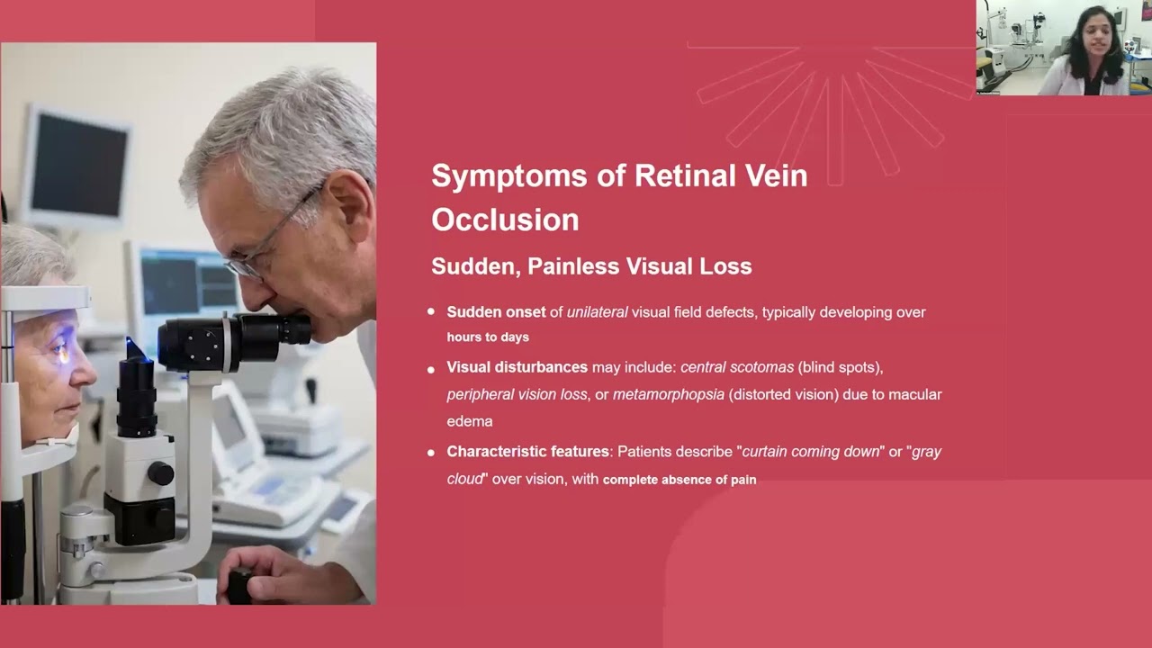
- The primary symptom is sudden, painless vision loss, which can range from mild blurriness to severe vision impairment.
- Patients may experience visual disturbances like central scotomas, peripheral vision loss, or metamorphopsia (distorted vision) if macular edema is present.
- CRVO typically presents with diffuse retinal hemorrhages, venous dilation and tortuosity, cotton wool spots, and sometimes optic disc swelling.
- The 'blood and thunder' appearance is a characteristic, though not universal, description of the fundus in CRVO.
- Distinguishing between ischemic and non-ischemic CRVO is crucial for prognosis and management.
- A relative afferent pupillary defect (RAPD) is a key clinical sign suggestive of significant retinal ischemia and is more prominent in ischemic CRVO.
- BRVO affects a specific sector or quadrant of the retina, typically occurring at arteriovenous crossings, most commonly in the superotemporal quadrant.
- Symptoms and signs are localized to the affected quadrant, including sectoral hemorrhages, venous dilation, and potential macular edema.
- Collaterals are dilated pre-existing venous channels that form to bypass the obstruction, improving venous outflow and often indicating chronicity rather than active disease.
- Collaterals are a favorable sign and should generally be observed, not treated with laser or anti-VEGF therapy.
- Ophthalmic examination includes visual acuity, slit lamp examination (checking for neovascularization of the iris/angles), gonioscopy, and fundus examination.
- Optical Coherence Tomography (OCT) is essential for quantifying macular edema, assessing retinal thickness, and identifying cystoid spaces or subretinal fluid.
- Fundus Fluorescein Angiography (FFA) is used to assess capillary perfusion, differentiate ischemic from non-ischemic RVO, and detect neovascularization.
- OCT Angiography (OCTA) provides non-invasive visualization of retinal vasculature and can detect macular ischemia and neovascularization, though it doesn't show leakage.
- The primary goals of management are visual preservation, treatment of macular edema, prevention of neovascularization, and control of systemic risk factors.
- Anti-VEGF therapy (e.g., ranibizumab, aflibercept) is the first-line treatment for RVO-related macular edema.
- Steroid implants (e.g., dexamethasone) can be used as a second-line therapy or in combination with anti-VEGF for persistent edema.
- Pan-retinal photocoagulation (PRP) is the standard treatment for neovascularization to induce regression of abnormal vessels and prevent complications like vitreous hemorrhage or neovascular glaucoma.
- Common complications include macular edema, retinal ischemia, and neovascularization (of the disc, elsewhere, or iris).
- Neovascular glaucoma is a severe, vision-threatening emergency resulting from neovascularization in the anterior chamber angle, leading to rapid IOP elevation.
- Regular follow-up is essential, including monthly visits initially, to monitor for disease progression, recurrence, and the development of complications.
- Patient education on the chronic nature of the disease, adherence to treatment, and realistic expectations is vital for successful long-term management.
Key takeaways
- Retinal vein occlusions are significant vascular events causing vision loss, with hypertension being the most common risk factor.
- Differentiating between ischemic and non-ischemic CRVO, often indicated by RAPD, is critical for prognosis and treatment decisions.
- Macular edema is the primary cause of vision loss in RVOs and is effectively treated with anti-VEGF therapy.
- Neovascularization is a serious complication of ischemic RVOs, requiring prompt treatment with pan-retinal photocoagulation to prevent severe vision loss.
- A comprehensive systemic evaluation is mandatory, especially in young patients or those with bilateral RVOs, to identify underlying pro-thrombotic or inflammatory conditions.
- Collateral vessels are a sign of the eye's adaptation and generally do not require treatment, but close follow-up is still necessary.
- Early recognition, prompt treatment, and consistent long-term follow-up are paramount for optimizing visual outcomes in patients with RVO.
Key terms
Test your understanding
- What are the three components of Virchow's triad that contribute to the pathophysiology of retinal vein occlusions?
- How does the presence of a relative afferent pupillary defect (RAPD) help differentiate between ischemic and non-ischemic Central Retinal Vein Occlusions (CRVOs)?
- What is the primary mechanism by which anti-VEGF therapy improves vision in patients with RVO-related macular edema?
- Why is a comprehensive systemic evaluation particularly important for young patients diagnosed with a retinal vein occlusion?
- What is the role of collaterals in the management of Branch Retinal Vein Occlusions (BRVOs), and how are they typically managed?