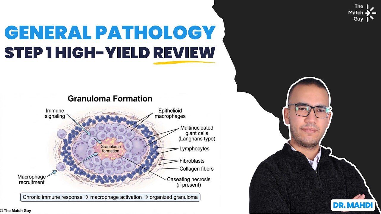
Step 1 General Pathology USMLE High-Yield Review
USMLE Tutoring - The Match Guy
Overview
This video provides a high-yield review of general pathology concepts crucial for USMLE Step 1 preparation. It covers cellular adaptations like hypertrophy, hyperplasia, metaplasia, and atrophy, explaining their mechanisms and clinical implications. The session also delves into cell death, differentiating between apoptosis and necrosis, and exploring their pathways and causes. Finally, it discusses various types of necrosis (coagulative, liquefactive, caseous, fat, gangrenous) and the pathogenesis of amyloidosis, linking these concepts to specific diseases and clinical scenarios. The presenter uses a question-based approach to reinforce learning and highlight key associations.
Save this permanently with flashcards, quizzes, and AI chat
Chapters
- Hypertrophy is an increase in cell size, often seen in permanent cells like cardiac muscle, in response to stress (e.g., hypertension leading to left ventricular thickening).
- Hyperplasia is an increase in cell number, occurring in labile or stable cells that can divide (e.g., smooth muscle cells in severe hypertension causing 'onion skin' appearance in arterioles).
- Metaplasia is a reversible change where one differentiated cell type is replaced by another, usually in response to chronic stress (e.g., columnar to squamous epithelium in smokers' airways, increasing cancer risk).
- Atrophy is a decrease in cell size and number, often due to decreased hormonal stimulation, disuse, or denervation (e.g., testicular atrophy from anabolic steroid use).
- Apoptosis is programmed cell death, a 'polite' process characterized by cell shrinkage, nuclear fragmentation, and no inflammation, essential for normal development and tissue homeostasis.
- Necrosis is accidental cell death, a 'nasty' process involving cell swelling, inflammation, and disorganized breakdown, typically caused by injury or toxins.
- The intrinsic pathway of apoptosis is triggered by internal cellular damage, involving the release of cytochrome C from mitochondria and activation of caspases.
- The extrinsic pathway of apoptosis is triggered by external signals binding to death receptors (e.g., Fas ligand binding Fas receptor), also leading to caspase activation.
- Cytotoxic T cells and NK cells induce apoptosis via the perforin-granzyme pathway, creating pores and delivering granzymes to activate caspases.
- Coagulative necrosis, characterized by preserved tissue architecture, occurs in most ischemic injuries (e.g., heart attack).
- Liquefactive necrosis, resulting in a liquid-filled cavity, is seen in brain ischemia (due to microglial enzymes) and abscesses (due to bacterial enzymes).
- Caseous necrosis, with a cheesy appearance, is characteristic of tuberculosis.
- Fat necrosis occurs when fatty acids bind calcium, forming 'saponification,' often seen in acute pancreatitis and breast trauma.
- Gangrenous necrosis is a clinical term for significant tissue loss, often appearing mummified (dry gangrene) or wet (superinfected) in extremities or the bowel.
- Amyloidosis is the extracellular deposition of misfolded proteins (amyloid) that the body cannot clear, leading to organ dysfunction.
- AL amyloidosis is associated with plasma cell malignancies like multiple myeloma, where excess immunoglobulin light chains deposit.
- AA amyloidosis occurs in chronic inflammatory states (e.g., rheumatoid arthritis, lupus).
- Beta-2 microglobulin amyloidosis is seen in patients with end-stage renal disease on dialysis, potentially causing carpal tunnel syndrome.
- Transthyretin amyloidosis can be age-related or familial (mutant form), affecting the heart and nerves.
- Multiple myeloma is characterized by CRAB symptoms: hypercalcemia, renal failure, anemia, and bone lesions.
- The immune system eliminates excess activated T cells after infection via apoptosis, a process crucial for restoring lymphocyte homeostasis.
- Granulomatous inflammation involves the formation of granulomas, collections of macrophages (epithelioid histiocytes) and other immune cells, often seen in response to persistent pathogens like TB.
- The development of granulomas involves macrophages engulfing pathogens, presenting antigens to CD4+ T cells, which differentiate into T helper 1 (Th1) cells.
- Th1 cells release interferon-gamma (IFN-γ), which activates macrophages to become epithelioid histiocytes, forming granulomas.
- Inhibition of IFN-γ can lead to impaired granuloma formation and progression from latent to active tuberculosis.
- Granulomas can be caseating (with central necrosis, typical of TB) or non-caseating (without necrosis, seen in sarcoidosis or Crohn's disease).
Key takeaways
- Cellular adaptations like hypertrophy and hyperplasia allow tissues to respond to stress, but metaplasia and atrophy can indicate pre-cancerous changes or tissue degradation.
- Apoptosis is a controlled, programmed cell death essential for development and tissue maintenance, distinct from the inflammatory, accidental cell death of necrosis.
- The specific gross appearance of necrotic tissue (coagulative, liquefactive, caseous, fat, gangrenous) provides vital diagnostic clues about the underlying cause.
- Amyloidosis results from the accumulation of misfolded proteins and can lead to severe organ damage, with different protein types linked to specific underlying conditions like multiple myeloma or chronic inflammation.
- Effective control of chronic infections like tuberculosis relies on a robust granulomatous inflammatory response mediated by T helper 1 cells and interferon-gamma.
- Understanding the mechanisms of cell adaptation, death, and immune response is fundamental to diagnosing and managing a wide range of pathologies tested on Step 1.
Key terms
Test your understanding
- How does hypertrophy differ from hyperplasia in terms of cellular response to stress, and what are examples of each?
- What are the key histological and clinical differences between apoptosis and necrosis?
- Describe the different types of necrosis and the specific conditions or injuries typically associated with each.
- What is amyloidosis, and how do the different types (e.g., AL, AA, beta-2 microglobulin) relate to specific diseases or patient populations?
- Explain the role of T helper 1 cells and interferon-gamma in the formation of granulomas, and how their dysfunction can lead to active tuberculosis?