9:40
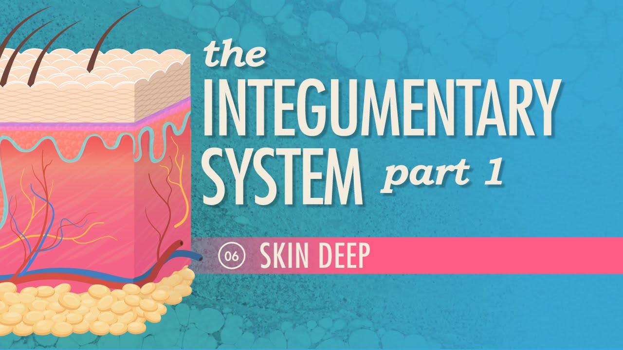
The Integumentary System, Part 1 - Skin Deep: Crash Course Anatomy & Physiology #6
CrashCourse
Overview
This video introduces the integumentary system, focusing on the skin as its primary organ. It details the skin's crucial functions, including protection, temperature regulation, and sensory perception. The video breaks down the skin into its three main layers—epidermis, dermis, and hypodermis—explaining the distinct cell types and structures within each. It highlights how these layers work together to maintain bodily integrity and interact with the external environment, emphasizing the importance of skin health and protection from damage.
How was this?
Save this permanently with flashcards, quizzes, and AI chat
Chapters
- The skin is the body's largest organ, performing vital functions like protection, fluid balance, and vitamin D synthesis.
- It contains nerve endings for sensation and structures like sweat glands and blood vessels for temperature regulation and emotional expression.
- The integumentary system includes the skin, hair, nails, and glands, all working together.
- Skin damage can lead to rapid complications but also exhibits quick healing capabilities.
Understanding the skin's role as a major organ highlights its importance in overall health and its complex functions beyond just covering the body.
The skin's ability to regulate temperature is demonstrated through sweating and flushing, which are visible responses to environmental conditions or emotional states.
- The epidermis is the outermost, visible layer, composed of stratified squamous epithelial tissue.
- Keratinocytes produce keratin, a tough protein that provides waterproofing and durability to the skin, hair, and nails.
- Melanocytes produce melanin, the pigment responsible for skin color, with variations in color determined by melanin amount, not cell number.
- Dendritic (Langerhans) cells act as immune cells, engulfing invaders, while Merkel cells at the base detect touch.
The epidermis is the body's first line of defense, and understanding its cellular composition explains how it protects against physical damage, UV radiation, and pathogens.
Tattoos are permanent because the ink is deposited below the epidermis, into the dermis, illustrating the epidermal layer's role as a barrier that sheds cells over time.
- Thick skin (palms, soles) has five epidermal layers, while thin skin has four.
- The stratum corneum (horny layer) is the outermost layer of dead keratinocytes, providing basic protection.
- The stratum lucidum (clear layer) is found only in thick skin.
- Deeper layers like stratum granulosum, stratum spinosum, and stratum basale contain living keratinocytes that mature and move upwards, with the stratum basale being the primary site of cell regeneration.
- The epidermis is avascular, relying on the dermis for nutrients, which contributes to cell death as they move away from the blood supply.
Knowing the specific layers and their functions explains how the epidermis regenerates and why certain areas of the skin are tougher than others, as well as the consequences of damage.
Stepping on a nail illustrates the progression through the epidermal layers: piercing the dead cells of the stratum corneum, potentially the stratum lucidum, then the living cells of the stratum granulosum and spinosum, and finally reaching the stratum basale.
- The dermis lies beneath the epidermis and contains collagen and elastin fibers for strength and elasticity.
- It is rich in capillaries, blood vessels, and nerve fibers responsible for sensation (temperature, pressure, pain).
- The dermis houses hair follicles, oil glands, and sweat glands.
- The papillary layer, the upper part, contains dermal papillae that form friction ridges (fingerprints).
- The reticular layer, the deeper and thicker part, contains dense irregular connective tissue and houses most of the dermis's structures.
The dermis is the powerhouse of the skin, containing the structures necessary for sensation, nourishment, and support, making its integrity crucial for overall skin function.
When a cut is deep enough to cause bleeding and pain, it signifies that the epidermis has been penetrated and the dermis has been damaged, highlighting the dermis's vascularity and nerve supply.
- The hypodermis, or subcutis, is the deepest layer of the skin.
- It is primarily composed of adipose (fat) tissue.
- Functions include insulation, energy storage, shock absorption, and anchoring the skin to underlying structures.
This layer provides essential cushioning and insulation, demonstrating how fat storage contributes to thermoregulation and protection of deeper tissues.
The hypodermis is where the majority of the body's fat is stored, acting as a protective cushion for organs and helping to maintain body temperature.
Key takeaways
- The skin is a dynamic, multi-functional organ essential for survival, not just a passive covering.
- The three layers of the skin (epidermis, dermis, hypodermis) have distinct structures and roles that contribute to overall skin integrity.
- The epidermis constantly regenerates, with specialized cells like keratinocytes and melanocytes determining its protective and pigmentary functions.
- The dermis is the vascularized and innervated layer responsible for sensation, support, and housing accessory structures like glands and follicles.
- Damage to the skin, especially the dermis, can have significant consequences due to its critical role in sensation and support.
- Protecting the skin from damage, particularly from UV radiation, is vital for preventing premature aging and skin cancer.
Key terms
Integumentary SystemEpidermisDermisHypodermis (Subcutis)KeratinocytesKeratinMelanocytesMelaninDendritic Cells (Langerhans Cells)Merkel CellsStratum CorneumStratum BasaleDermal PapillaeAdipose Tissue
Test your understanding
- What are the three main layers of the skin and what is the primary function of each?
- How do keratinocytes and melanocytes contribute to the structure and appearance of the epidermis?
- Why is the epidermis avascular, and what are the implications of this for cell survival and regeneration?
- What key structures are found within the dermis, and how do they contribute to the skin's functions?
- Explain the role of the hypodermis in insulation and protection.