5:53
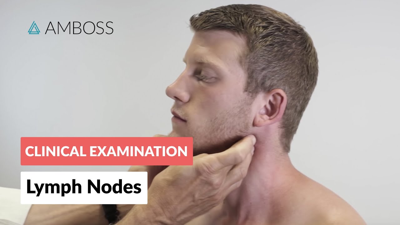
Examination of the Lymph Nodes - Clinical Examination
AMBOSS: Medical Knowledge Distilled
Overview
This video details the systematic clinical examination of lymph nodes, a crucial part of assessing for various health conditions. It emphasizes a head-to-toe approach, covering key lymph node regions: head and neck, axilla, and inguinal area. The examination involves both inspection and palpation, with specific techniques described for each region to identify abnormalities like enlargement, tenderness, or unusual consistency. The video also briefly touches upon the examination of the spleen as it relates to lymphatic system assessment.
How was this?
Save this permanently with flashcards, quizzes, and AI chat
Chapters
- Clinical examination of lymph nodes involves systematic inspection and palpation.
- Key lymph node regions to examine are the head and neck, axilla, and inguinal area.
- The examination typically proceeds from cranial (head) to caudal (inguinal).
A systematic approach ensures all clinically relevant lymph node stations are assessed, increasing the likelihood of detecting abnormalities that could indicate disease.
- About one-third of all lymph nodes are in the head and neck, making them easily palpable.
- Specific nodes to palpate include sub-occipital, pre/retro-auricular, submandibular, sub-mental, posterior triangle, and those near the internal jugular vein.
- Supraclavicular lymph nodes are critical to check, as their enlargement can signal malignancies, including abdominal cancers like gastric cancer (Virchow's node).
The head and neck region contains numerous superficial lymph nodes, making them accessible for early detection of infections or cancers that may have spread from other areas.
Enlargement of the Virchow node in the left supraclavicular fossa is a significant finding often associated with abdominal tumors that have metastasized via the lymphatic system.
- Axillary lymph nodes are divided into pectoral (anterior), subscapular (posterior), brachial (lateral), central, and apical groups.
- These groups drain lymphatic fluid from the chest, chest wall, and arms.
- Palpation is best performed with the arm relaxed to differentiate nodes from muscles.
- The examiner palpates the pectoral, central, posterior, and brachial groups in sequence.
Examining axillary lymph nodes is vital for detecting issues related to the breast, upper limbs, and chest wall, as these nodes are primary drainage sites for these areas.
The examiner palpates the pectoral group behind the pectoralis major muscle, then the central group at the axilla's base, followed by the posterior and brachial groups.
- Superficial inguinal lymph nodes are divided into horizontal and vertical groups.
- The horizontal group lies below the inguinal ligament and drains the external genitalia, trunk, and lower back.
- The vertical group is near the great saphenous vein and drains the lower extremity.
- Examination is best done with the patient lying down.
Assessment of the inguinal lymph nodes helps identify potential infections or malignancies in the lower extremities, perineal region, and lower abdomen.
The horizontal group is palpated parallel to the inguinal ligament, while the vertical group is assessed adjacent to the great saphenous vein.
- The spleen, a lymphatic organ, should be considered during lymph node assessment.
- Splenomegaly (enlarged spleen) can indicate systemic inflammation or malignancy.
- A healthy spleen is usually not palpable; an enlarged spleen can be felt under the left costal margin during inspiration.
- Palpation technique involves lifting the patient's left flank to aid detection.
The spleen's involvement in lymphatic and immune functions means its size can reflect systemic diseases, complementing the findings from lymph node examination.
During inspiration, the inferior edge of a pathologically enlarged spleen descends and may be felt by the examiner's fingertips below the left costal margin.
Key takeaways
- A systematic, head-to-toe examination of lymph node stations (head/neck, axilla, inguinal) is essential for clinical assessment.
- Enlarged, tender, or unusually firm lymph nodes, especially when fused into conglomerates, are significant findings requiring further investigation.
- The location of enlarged lymph nodes provides clues to the origin of potential disease, such as Virchow's node indicating abdominal malignancy.
- Proper technique, including patient relaxation and specific palpation methods for each region, improves the accuracy of lymph node examination.
- Assessing the spleen alongside lymph nodes offers a broader view of potential systemic inflammatory or malignant conditions.
- Understanding the lymphatic drainage patterns of different body areas is crucial for interpreting lymph node findings.
Key terms
Lymph nodesPalpationInspectionAxillaInguinal areaSupraclavicular lymph nodesVirchow's nodeConglomeratesSplenomegaly
Test your understanding
- What are the three primary lymph node regions that should be systematically examined?
- Why is palpating the supraclavicular lymph nodes particularly important?
- How does the technique for examining axillary lymph nodes differ from inguinal lymph node examination?
- What characteristics of a palpable lymph node (e.g., consistency, mobility) should be noted, and why?
- How can the examination of the spleen contribute to the overall assessment of the lymphatic system?