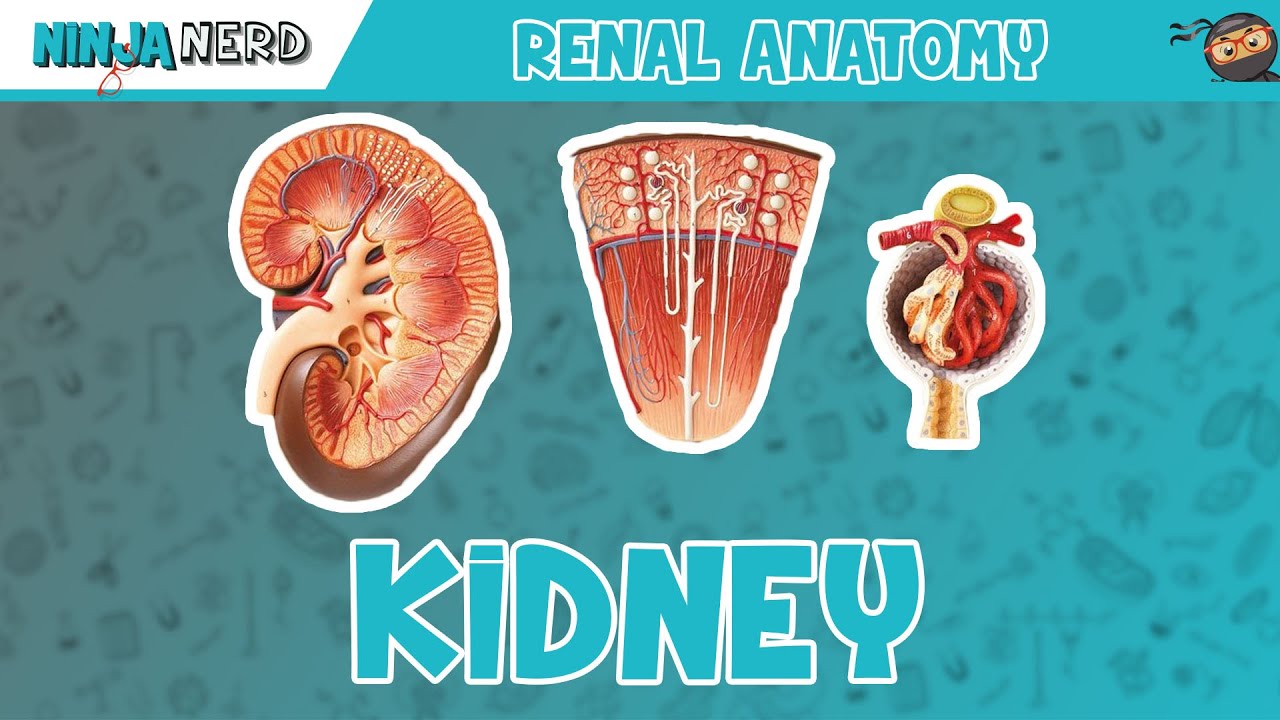
Renal | Kidney Anatomy Model
Ninja Nerd
Overview
This video provides a detailed anatomical overview of the kidney, covering its external structures, internal regions, and the intricate blood supply system. It explains the layers of tissue surrounding the kidney, the entry and exit points for vessels and the ureter (hilus and sinus), and delineates the cortex and medulla. The video then traces the path of blood flow through various arteries and veins within the kidney and describes the functional unit, the nephron, detailing its components from the renal corpuscle to the collecting duct. Finally, it elaborates on the renal corpuscle, including the glomerulus, Bowman's capsule, and the juxtaglomerular apparatus, highlighting their roles in filtration and blood pressure regulation.
Save this permanently with flashcards, quizzes, and AI chat
Chapters
- The kidneys are bean-shaped organs located in the abdominal cavity.
- The renal capsule, a layer of loose connective tissue, directly surrounds the kidney to prevent infections.
- External to the renal capsule are the perirenal fat capsule (adipose tissue for cushioning) and the renal fascia (dense fibrous tissue anchoring the kidney).
- The renal hilus is a slit-like indentation where the renal artery, renal vein, and ureter enter or exit.
- The renal sinus is the internal space containing the renal pelvis, vessels, and other structures.
- The outer region of the kidney, appearing granulated and beige, is the renal cortex.
- The inner region, dark and striated, is the renal medulla, which is organized into cone-shaped structures called renal pyramids.
- Renal columns are inward extensions of the cortical tissue that dip down between the renal pyramids.
- The tip of each renal pyramid is called the renal papilla, which is where urine collects.
- Each renal papilla drains urine into a minor calyx.
- Multiple minor calyces merge to form a larger major calyx.
- Several major calyces converge to form the renal pelvis, a funnel-shaped structure.
- The renal pelvis exits the kidney at the hilus and becomes the ureter, which carries urine to the bladder.
- Blood enters the kidney via the renal artery, which branches from the abdominal aorta.
- Inside the kidney, the renal artery divides into segmental arteries.
- Segmental arteries lead to interlobar arteries, which run between the renal lobes (pyramid + surrounding cortex).
- Interlobar arteries branch into arcuate arteries at the corticomedullary junction.
- Arcuate arteries give rise to cortical radiate arteries, which further branch into afferent arterioles supplying the nephrons.
- Blood leaving the nephrons collects in peritubular capillaries and the vasa recta.
- These capillaries drain into cortical radiate veins.
- Cortical radiate veins merge to form arcuate veins.
- Arcuate veins drain into interlobar veins, which run between the lobes.
- Finally, interlobar veins converge to form the renal vein, which exits the kidney.
- A nephron consists of the renal corpuscle (glomerulus and Bowman's capsule) and the renal tubule (proximal convoluted tubule, loop of Henle, distal convoluted tubule).
- The renal corpuscle filters blood plasma, with the filtrate entering Bowman's capsule.
- The proximal convoluted tubule reabsorbs essential nutrients and solutes.
- The loop of Henle (descending and ascending limbs) plays a role in concentrating urine.
- The distal convoluted tubule further modifies the filtrate through secretion and reabsorption.
- The collecting duct receives filtrate from multiple distal convoluted tubules and transports it towards the renal papilla.
- The renal corpuscle contains the glomerulus (a capillary tuft) surrounded by Bowman's capsule.
- The afferent arteriole brings blood to the glomerulus, and the efferent arteriole drains it.
- Podocytes, specialized cells forming the visceral layer of Bowman's capsule, cling to the glomerular capillaries.
- The juxtaglomerular apparatus includes JG (juxtaglomerular) cells in the afferent arteriole (baroreceptors) and macula densa cells in the distal convoluted tubule (chemoreceptors).
- This apparatus regulates blood pressure by secreting renin and monitors filtrate composition.
Key takeaways
- The kidney is protected by multiple layers of connective tissue, including the renal capsule, perirenal fat capsule, and renal fascia.
- The kidney's internal structure is divided into the outer cortex and the inner medulla, which contains renal pyramids.
- Urine travels sequentially through minor calyces, major calyces, the renal pelvis, and finally exits via the ureter.
- The intricate network of arteries (renal, segmental, interlobar, arcuate, cortical radiate, afferent) and veins (efferent, peritubular, vasa recta, cortical radiate, arcuate, interlobar, renal) supplies and drains blood from the nephrons.
- The nephron, composed of the renal corpuscle and renal tubule, is the functional unit responsible for filtering blood and producing urine.
- The renal corpuscle, containing the glomerulus and Bowman's capsule, is where filtration begins.
- The juxtaglomerular apparatus, formed by JG cells and macula densa cells, is crucial for regulating blood pressure and monitoring filtrate.
Key terms
Test your understanding
- What are the three layers of tissue surrounding the kidney, and what is the primary function of each?
- How does urine flow from the renal papilla to the ureter, and what structures are involved in this pathway?
- Trace the path of blood flow from the renal artery to the afferent arteriole, naming the key arterial branches.
- Describe the main components of a nephron and the general role of each component in urine formation.
- What is the juxtaglomerular apparatus, and how do its components (JG cells and macula densa cells) contribute to regulating blood pressure and filtrate?