The Nervous System - PART 1
Atty Gab
Overview
This video introduces the anatomy of the human nervous system, dividing it into the central nervous system (brain and spinal cord) and the peripheral nervous system (sensory and motor). It details the functions of the sympathetic and parasympathetic nervous systems, explaining their roles in 'fight or flight' and 'rest and digest' responses, respectively, and their impact on vital signs and bodily functions. The video then explores the four main divisions of the brain: the cerebrum (with its white and gray matter, and lobes like frontal, temporal, parietal, and occipital), the diencephalon (housing the hypothalamus, thalamus, epithalamus, and pituitary gland), the cerebellum (responsible for balance), and the brainstem (including the pons, medulla oblongata, and midbrain), which controls vital autonomic functions and reflexes.
Save this permanently with flashcards, quizzes, and AI chat
Chapters
- The nervous system is divided into the Central Nervous System (CNS) and the Peripheral Nervous System (PNS).
- The CNS consists of the brain and spinal cord.
- The PNS is further divided into sensory (afferent) and motor (efferent) pathways.
- Sensory pathways transmit information from stimuli to the brain, while motor pathways transmit responses from the brain to muscles.
- The motor division of the PNS includes voluntary (somatic) and involuntary (autonomic) responses.
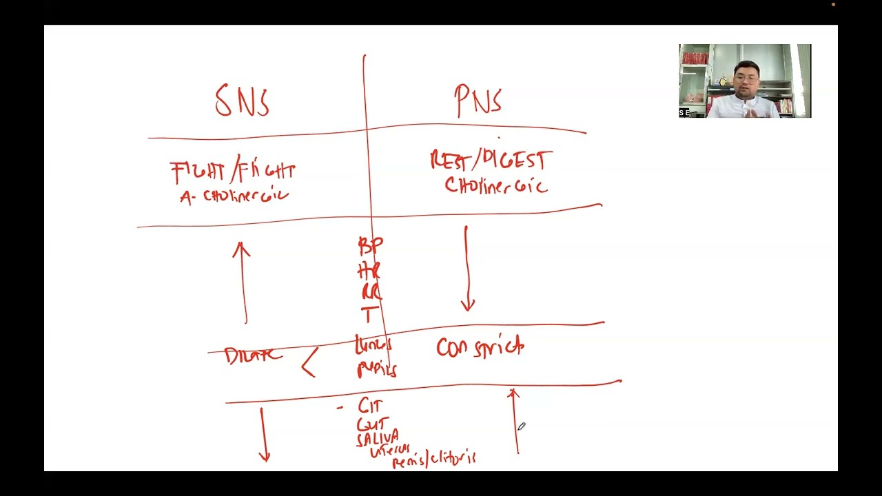
- The autonomic nervous system is further divided into the sympathetic (fight or flight) and parasympathetic (rest and digest) systems.
- Sympathetic activation increases heart rate, blood pressure, and dilates pupils and lungs, preparing the body for stress.
- Parasympathetic activation slows heart rate, constricts pupils and lungs, and promotes digestion and rest.
- Understanding these opposing functions is key to predicting medication side effects (e.g., cholinergic vs. anticholinergic).
- The cerebrum, the largest part of the brain, is composed of white matter (connecting hemispheres) and gray matter (housing lobes).
- The frontal lobe is associated with personality, attention, speech (Broca's area), and thinking.
- The temporal lobe processes auditory information and contains Wernicke's area for language comprehension.
- The parietal lobe processes sensory information like pressure and proprioception (body position).
- The occipital lobe is dedicated to visual processing.
- The diencephalon includes the hypothalamus (appetite, temperature, thirst, love, puberty), thalamus (sensory relay station), and epithalamus (pineal body for melatonin, choroid plexus for CSF production).
- The cerebellum is crucial for maintaining balance and equilibrium, resembling a cauliflower in shape.
- The brainstem comprises the pons (respiration depth), medulla oblongata (respiration rate, heart rate), and midbrain (auditory and visual reflexes).
- The reticular formation in the brainstem regulates the sleep-wake cycle.
- Cerebrospinal fluid (CSF) is produced by the choroid plexus within the epithalamus.
- CSF circulates through ventricles and the subarachnoid space, cushioning the brain and protecting it from impact.
- The normal volume of CSF in circulation is about 150 ml; excess can lead to hydrocephalus (in infants) or increased intracranial pressure (in older children/adults).
- The skull sutures allow for brain growth in infants, preventing increased intracranial pressure.
- Signs of increased intracranial pressure include elevated BP, decreased heart rate and respiration, and increased temperature (the 'rock and roll' sign).
Key takeaways
- The nervous system's division into CNS and PNS provides a framework for understanding neural pathways.
- The opposing actions of the sympathetic and parasympathetic systems are essential for maintaining homeostasis under varying conditions.
- Specific brain lobes are specialized for distinct functions, enabling complex cognitive processes and sensory perception.
- The diencephalon, cerebellum, and brainstem control vital autonomic functions, balance, and consciousness.
- Cerebrospinal fluid is critical for protecting the brain and maintaining intracranial pressure, with imbalances indicating potential pathology.
- Understanding the relationship between drug effects and autonomic nervous system activation is crucial in pharmacology.
- The brain's structure, from its lobes to its protective CSF, is intricately designed for optimal function and safety.
Key terms
Test your understanding
- How do the central and peripheral nervous systems work together to process sensory information and generate a response?
- What are the primary physiological differences between sympathetic and parasympathetic nervous system activation, and why is this distinction important for understanding medication effects?
- Describe the main functions of the frontal, temporal, parietal, and occipital lobes of the cerebrum.
- Explain the role of the hypothalamus and the thalamus within the diencephalon.
- What is the function of cerebrospinal fluid, and what are the potential consequences of its abnormal accumulation or pressure?
- How do the pons and medulla oblongata in the brainstem regulate breathing and heart rate?